Learning Outcomes:
i. Students will understand the principles of light microscopy and electron microscopy.
ii. They will be able to distinguish between the functionalities and capabilities of both types of microscopy.
iii. Students will recognize the impact of microscopy on our understanding of cellular structures and functions.
Summary of Lesson:
Microscopes are the tools that have allowed us to enter and explore the tiny universe that was once invisible to the naked eye. This lesson unveils the intricacies of light and electron microscopy, the technologies that have greatly expanded our understanding of the microscopic world.
Content:
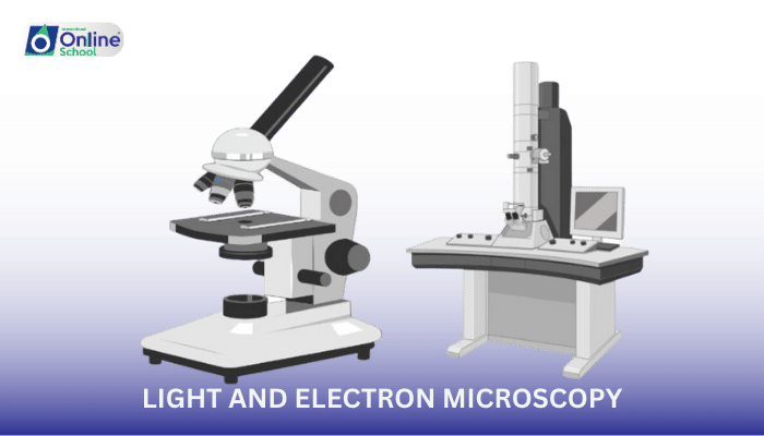
i. The Essence of Light Microscopy: Light microscopy, one of the oldest scientific instruments, uses visible light to illuminate and magnify specimens. It has lenses that bend light to magnify images of small objects, allowing us to see individual cells and even some larger organelles within them.
ii. The Power of Electron Microscopy: Electron microscopy, which emerged in the 20th century, uses a beam of electrons instead of light to create an image. It has a much higher resolution than light microscopy, revealing the ultrastructure of cells, viruses, and proteins in fine detail.
iii. Comparing Light and Electron Microscopy: Magnification and Resolution: Light microscopes can magnify up to about 1000 times, while electron microscopes can magnify up to two million times.
iv.Sample Preparation: Specimens for light microscopy can often be viewed live or with minimal preparation, whereas electron microscopy requires samples to be fixed, dehydrated, and sometimes sliced into ultra-thin sections.
v. Imaging Conditions: Light microscopy can be performed in natural conditions, while electron microscopy requires a vacuum as electrons are easily scattered by air molecules.
vi. Cost and Accessibility: Light microscopes are more common and less expensive than electron microscopes, which are larger and require more maintenance.
List of Important Questions for Self-Study:
i. What is the basic principle behind light microscopy?
ii. How does electron microscopy differ from light microscopy in terms of image creation?
iii. What are the limitations of light microscopy in observing cellular structures?
iv. Why can electron microscopy achieve higher resolution than light microscopy?
v. How does the requirement for a vacuum in electron microscopy affect sample observation?
vi. Why can't living specimens be observed using electron microscopy?
vii. What are some of the practical applications of light and electron microscopy in biology?
viii. How has electron microscopy enhanced our understanding of diseases at the cellular level?
ix. Why is light microscopy still widely used despite the higher resolution of electron microscopy?
x. Can you think of a scenario where you would prefer to use light microscopy over electron microscopy?
Important Terminologies Used in Lesson:
i. Light Microscopy: A technique in which light is passed through a specimen to an objective lens, typically used for lower magnification of samples.
ii. Electron Microscopy: A technique that uses a beam of accelerated electrons as a source of illumination, providing higher resolution images of biological samples.
iii. Magnification: The process of enlarging the appearance of an object via optical instruments.
iv. Resolution: The ability of a microscope to distinguish two adjacent points as separate and distinct.
v. Vacuum: A space devoid of matter, which is required for electron microscopy to prevent scattering of the electron beam.
vi. Sample Preparation: The process of treating a specimen to be viewed under a microscope, which can include fixation, sectioning, and staining.
